Scientific

anja_july_29th_lecture_at_skeri

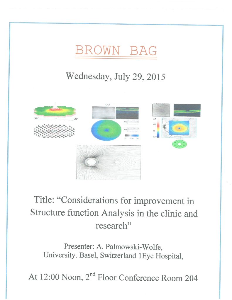
Considerations for Improvement in Structure Function Analysis in the Clinic and Research

Abstract: Recent imaging techniques with the OCT give fast and easy information on retinal anatomy. The question arises whether this examination renders functional tests unnecessary. Clinical examples will be given to show that these tests complement one another rather than substitute one another. Can segmented macular ganglion cell-inner plexiform layer (GCIPL) analysis further increase structure function correlation? How do different OCT brands (Spectralis and Cirrus) compare to one another and what impact do these differences have on structure function analysis. In regard to functional tests: Can visual field analysis be improved to better be able to compare structure to function? Can we improve our mfERG analysis to this regard.

prof_anja_wolfe_july_23_bb

Oculodynamic MRI– Possible Clinical Applications

Abstrat:   Oculodynamic MRI recordings can be obtained as an addition to routine MRI recordings in 30 additional seconds. I will show examples of these eye movements, correlate them to clinical findings and expand on how this may help in the treatment of patients. For instance, presence or absence of impingement can be seen dynamically and treatment of trauma or tumors may be planed accordingly. 3-D visualization demonstrates bulbar and global EOM layers. Orbital nerves and vascular structures can be analyzed using special positions of the clipping plane. Both parts of the lacrimal gland can be visualized in detail. In TAO the marked enlargement of the lacrimal gland, the EOM and the orbital fat can be appreciated in 3D. Thus odMRI and detailed 3D rendering of the orbit offer exciting new diagnostic and therapeutic potential.